| HOME | SPINAL CONDITIONS | PROCEDURES | CLINICS | BACKGROUND | CONTACT | MEDICO-LEGAL |
Procedures Performed |
Procedure ListClick on a heading below to find out more. Nerve Root BlocksCaudal EpiduralFacet Joint InjectionsFacet Joint CoblationLumbar DiscectomySpinal Decompression/LaminectomyPercutaneous NucleoplastyCOFLEX Ligament/WALLIS LigamentXLIF® ProcedureDynesisLumbar FusionLumbar Disc ReplacementCervical DiscectomyPosterior Cervical Decompression/StabilisationSpinal Surgery for TraumaPosterior Correction and Fusion for ScoliosisAnterior Correction of ScoliosisAnterior/Posterior Reconstruction of Tumours and Infection |
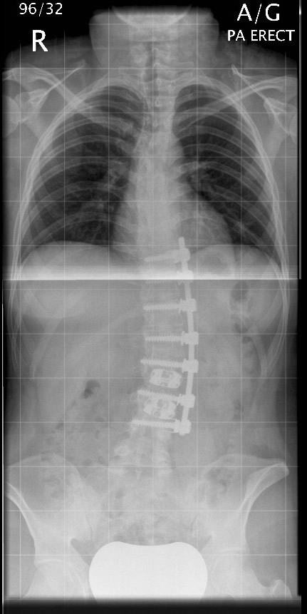
Nerve Root BlocksNerve root pain can result from irritation of the nerve from a disc prolapse or bony irritation. This presents as arm pain in neck, intercostal pain in the chest area and sciatica in the lower back. Nerve root blocks are carried out as day case procedures under x-ray screening. They can be used both as a diagnostic injection as well as therapeutic one and avoid the need for surgery. The diagnostic purpose is to show the treating surgeon precisely which nerve is causing the symptom. The injection allows local anaesthetic and steroid to be injected around the nerve to help calm the nerve down and decrease the patient’s pain. This can give a permanent solution to the nerve pain, although on occasion the pain can return. Caudal EpiduralA caudal epidural relates to the injection of steroid and local anaesthetic into the space that surrounds the nerves in the spinal canal. Nerves can be irritated from a disc prolapse or from bony irritation and an injection into the epidural space can reduce the inflammation around the nerve helping reduce the patient’s back and more specifically leg symptoms. Epidurals are very useful in patients who have spinal stenosis where bilateral symptoms occur. The procedure is carried out as a day case procedure and can be carried out under sedation if required. The injection is carried out through the sacral hiatus between the sacrum and the coccyx. Some people get occasional numbness into the legs but this wears off within a matter of a few hours. The injection can take four to six weeks to take full effect. It can be repeated depending on the longevity of the pain relief. There is a small risk of infection at the injection site as well as headache due to inadvertent puncture of the dura, which surrounds the nerve roots that retain fluid support to the nerve, this tends to settle by itself. Facet Joint InjectionsFacet joints are joints such as the hip or knee joints which are present in the posterior part of the spinal column. There is a pair at each level from the cervical down to the lumbar spine. These can undergo arthritic change and can lead on to pain in the back, buttocks and referred down into the legs but not normally passed the knee. Symptoms tend to be related to stiffness and pain especially when standing or leaning backwards. Facet joint injections are performed as a day case procedure using x-ray. Injection of local anaesthetic and steroid into either side of the spine into the facet joints is performed. Occasionally it is necessary to inject up to three facet joints on either side in one sitting. Facet joint injections can be diagnostic to help tell where the back pain can be coming from but also are therapeutic in the fact that they give pain relief. Facet Joint CoblationFacet Joint Coblation is aimed at providing longer term relief to facet pain. In patients who have had a good response to facet injections but the pain relief was only temporary, facet coblation aims at destroying the small pain fibres which give rise to pain from the facet joints. Facet joint injections are performed first as they carry a diagnostic purpose and give an idea whether facet coblation would be successful. Facet joint coblation is performed under sedation as a day case. The patient is allowed home later the same day. Lumbar DiscectomyThis procedure is used to treat such conditions such as disc herniation. The surgery is usually performed to relieve sciatica. Associated weakness or numbness can take several weeks to months to improve but 90% of patients find benefit in their leg pain reasonably quickly following the surgery. The surgery involves making a small incision approximately an inch or so over the disc and some bone and ligament is removed to gain access to the nerve that is irritated. The nerve is gently dissected off the disc prolapse and the disc material removed which allows the nerve to settle down. This procedure involves an average hospital stay of approximately three nights and mobilisation tends to occur within 24 hours of the procedure. If a drain is placed then it is removed 24 hours following the procedure. Prior to discharge, you will be independent including being able to go up and down stairs. It is advisable not to sit for prolonged periods following the surgery but sitting times can increase over the next few weeks and because of this we suggest avoiding driving for at least three weeks following the procedure. With regards to work, it depends on the patient’s employment but approximately six weeks is advisable and often a graded return to work is useful, especially for manual occupations. Physiotherapy is important and this will occur around the time of surgery and for the recuperation and rehabilitation period. Spinal Decompression/LaminectomyThe procedure is similar to discectomy but tends to involve removal of slightly more bone and a longer incision is made. Quite often muscles are dissected off the spine at both sides as the symptoms tend to be bilateral. Bone and ligament is removed to give freedom to the nerve which allows the nerve to recover. Again, any leg pain tends to respond to this treatment within a few days. Residual numbness/weakness can take several weeks or months to recuperate. Hospital stay for this procedure is roughly 3-4 days and following removal of the drain after 24 hours, mobilisation will commence with a physiotherapist. The patient will become more mobile daily and rehabilitation follows that of a discectomy procedure. Percutaneous NucleoplastyThis procedure is used to treat sciatica. A needle is placed into the disc using x-ray control. Once the needle is in the ideal position, a wire is passed down the needle and the disc is coblated, which removes disc material via vaporisation. This creates a cavity where a disc prolapse can collapse back into it, giving the nerve space. Benefits of this procedure are that no muscle dissection is performed and is done as a day case procedure under sedation. The risks of this procedure are small, with a small chance of damaging the nerve whilst passing the needle. This is minimised by the patient being sedated and their reaction can be monitored as the needle passes by the nerve. The benefit from this procedure can take a few weeks to present itself. There is roughly 80% benefit for leg pain following this treatment. Treatment is based on the individual disc prolapse on the MRI scan as some disc prolapses are amenable and other types are not, to this type of therapy. COFLEX Ligament/WALLIS LigamentThese implants are termed as interspinous process spacers and are used to enlarge the gap for the nerve as it comes out from the spinal canal. This can result in symptoms similar to sciatica. These implants can be used at the same time as decompressive surgery to give extra space for the nerves. Hospital stay with using these implants, is not lengthened simply to perform a decompression. XLIF® ProcedureThe XLIF® approach is a minimally disruptive procedure that allows the surgeon to have direct access to the intervertebral disc space and fuse the lumbar (low back) spine from the side of the body (lateral) as opposed to the front (anterior) or back (posterior). For more imformation see the XLIF website (Link). DynesisDynesis is an implant that provides a soft stabilisation. It involves inserting screws into the vertebrae above and below the affected disc for placing a plastic tube over a cord to link up the screws. This offloads the disc and is a treatment for specific types of disc pain. Whether the disc is suitable for this type of treatment depends on the disc height and also whether the disc has been confirmed to be the pain source on investigation. Lumbar Fusion
|
Design by Arcadia Software Limited. Copyright 2008-2010